ganglion cyst
Author:
Dr. Marc Mitnick
Reviewed by:
Medical Review Board
QUICK SUMMARY: WHAT IS A GANGLION CYST?
A ganglion cyst is a very common, benign (non-cancerous) lump that typically forms near joints or tendons. While they can appear anywhere, they frequently develop on the top of the foot (dorsal ganglion).
Essentially a fluid-filled sac, these "bible cysts" are often painless unless they press against a nerve or are irritated by footwear. Once properly diagnosed, if the cyst is not painful, no treatment is required.
IS A GANGLION A TUMOR?
It essentially is a benign tumor, or lump that can technically occur anywhere on the body but are most prevalent on the hands, but also very common on the feet. It is basically a sac filled with fluid that arises from either a joint (space between two bones) or from a tendon (structure that attaches a muscle into bone). There is a theory that it is nothing more than the result of herniation of a tendon, or synovium which makes up a joint.
A dorsal ganglion cyst which occurs on top of the foot is the most common location in the foot.
These cysts are also known as bible cysts or sometimes Gideon cysts because years ago the recommended treatment was to smash the growth with a book in order to break it up and even the poorest of families owned a bible.
The growth is more common in women with 70 percent of ganglions occurring in people between 20-40 years of age. On rare occasions they have been known to occur in children younger than 10 years of age. About 4% of them occur on the feet.
WHAT CAUSES A GANGLION CYST TO FORM
Ganglions usually occur due to a weakness in the covering of a tendon or joint space. Everyday trauma such as motion of a tendon over a bony prominence or pressure from a shoe on a tendon or joint can cause a weakness in the covering with a subsequent swelling of liquid into a confined space thus causing the “lump” to form.
symptoms of ganglion cyst
The growth may be moderately firm or soft, depending on the fluid content or whether the outer part of the sac bears calcification. The fluid itself is a colorless gel similar in appearance to k-y jelly. Pressure on the ganglion cyst itself rarely causes pain; any pain is from pressure to adjacent sites.
A diagnosis of a ganglion is usually made on clinical examination based on location of the growth and relevant history. An x-ray may be taken to rule out a bone spur as the aggravating factor behind the formation of the cyst. If there is any question as to the diagnosis an MRI or diagnostic ultrasound may be performed.
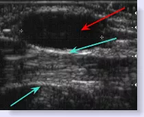
The picture below is that of a diagnostic ultrasound. The area in between the two blue arrows represents the tendon. Above the tendon is a black oval area (red arrow), which represents the ganglion. Since the cyst is made up of liquid it creates the least amount of “bounce” from the ultrasound waves and results in a black mass.
TREATMENT FOR GANGLION CYST
Once a mass is diagnosed as a ganglion cyst it may be left alone if it is not painful or causing problems with shoes. As stated previously sometimes these growths will spontaneously disappear. However, if there is any pain associated with the growth or the patient does not like the unsightly appearance, drainage is the simplest approach.
ganglion cyst aspiration
A large bore needle is inserted into the growth with local anesthesia and perhaps cortisone in an effort to burst and drain the growth. If the content of the ganglion is not too viscous this can be easily done; sometimes however, it is not possible to drain the cyst.
Once drained, a compression dressing is worn over the site for a period of time in an attempt to reduce recurrence. Having said that, there is a 70 percent recurrence rate with this form of treatment, but the upside is that the procedure can be repeated again since it is a relatively painless procedure.
ganglion cyst surgery
If the growth is a source of irritation for the patient then surgical excision is necessary keeping in mind that there is even a recurrence rate with this form of treatment. The problem with surgical excision is that sometimes the cyst will burst while attempting to remove it. Although the sac will still be present it becomes difficult to determine where the “stalk” or origin of the cyst came from. It is important to remove this stalk in an effort to prevent recurrence.
Aside from the possibility of recurrence, surgical complications are limited with stiffness in the surgical area being a primary complaint.
Below is a picture of a ganglion cyst that has just been identified prior to removal. It is the round glistening lesion located in between the blue arrows. The next picture is that of the cyst after its removal. In this instance the cyst was removed intact.
Surgical excision is an outpatient procedure with a quick recovery time frame.
There really is no preventative care for these cysts due to their spontaneous nature. It is no longer recommended to try and break them up by smashing them with a book as this can cause additional trauma to the area. Warm heat compresses might make the cyst and surrounding area feel a little more comfortable but will not rid you of the growth.
Once a diagnosis of ganglion cyst is made and more potentially dangerous growths have been ruled out, you can just leave the growth alone unless it bothers you.
Is a ganglion cyst a type of tumor?
Yes, it is a benign (non-cancerous) tumor or lump. It is a sac filled with a colorless, jelly-like gel that arises from a joint space or a tendon sheath.
Why is it sometimes called a "Bible Cyst"?
In the past, people would strike the cyst with a heavy book—often a family Bible—to rupture the sac. This is no longer recommended as it can damage surrounding foot structures.
Will a ganglion cyst go away on its own?
Yes, these cysts can spontaneously disappear without treatment. If the lump is not causing pain or shoe irritation, it is often best to simply monitor it.
What is the most common location for these on the foot?
The most common location is a dorsal ganglion, which occurs on the top of the foot.
Questions I Have Answered From Visitors
Question: Ganglion Cyst on the Bottom of Foot from Trauma
My husband was in an accident Dec of 09. He was driving a tractor trailer loaded with tree length wood. A women crossed the center line trying to avoid her when the truck rolled over. Trying to brake to stop on impact he took alot of force through his feet. He woke up face down in the passenger side of the truck.
They have surgically tryed to aspirate it twice with no luck. It is so large that he is having a hard time walking and his toes are turned up because he can not put pressure on it. We went to a specialist in Portland ME and he said he has never seen anything like that in his 30 years. They are in both feet but the right is much worse.
Our foot MD In Augusta ME said his team will be able to take it out but cutting through the bone on the top of his foot to get to it. Evidently because of the nerves and muscle it is easier to go through the top vs the bottom. The MRI's show that it is growing into the surrounding muscle and tissue ect.
This is 100% workers Comp and they agree after years of fighting finally. My husband is only 45 and I want him to have the best. I worry about the damage and complications after. I can not find anything on the internet relating to this type of surgery... None of the MD's doing this have ever done this before either... Though they "make" it sound easy. I would love your input or be able to get a resource of a MD who has done this.
Note: I have copies of MRI's for review.
Dr. Mitnick's Response
Hi Jennifer,
I am sure in the information you have found regarding ganglion cysts, every source will mention the fact that there is a better than fair chance of recurrence. That is the risk you take when you consent to having a ganglion surgically removed.
Having said that, based on your narrative, the risk/reward ratio favors your husband because he is in so much pain walking on it and previously attempted drainage of the cyst has not been successful.
The key to reducing the recurrence of a ganglion cyst is being able to dissect down to the root or stalk of the cyst and being able to excise the stalk. Anything short of that is subject to failure.
I know the cyst is located on the bottom of the foot and of course the common mantra in foot surgery is to avoid incisions on the bottom of the foot because of the potential chance of residual scarring of the incision which in itself may be painful to walk on.
So apparently in the way it is growing your doctor feels it is better to go through the top of the foot and through bone. I am assuming he is talking about one of the metatarsal bones, which are the long bones behind the toes.
Since visibility and being able to dissect to the base of the ganglion are so paramount, I am not so sure that going through the top of the foot and disrupting bone(s) is the best approach since I do not know if that will give them the best exposure to the base of the cyst. Of course they have examined your husband and seen the MRI; I have not.
On the surface at least I would think going through the bottom of the foot and taking the special precautions necessary when making this type of incision (scarring), would seem to be the better approach for any growth occurring on the bottom of the foot.
The main issue with going through bone is that they are going to have to saw through the bone and using a power saw to cut bone increases the chances of bursting the cyst, which then makes it impossible to properly excise the cyst.
I can only be of limited help here for the reasons I have previously stated. If there is real doubt on your part about the anticipated surgery, then why not get a second opinion in a bigger medical market like Boston for example. Barring any medical issues your husband may have, this would typically be an outpatient procedure, so it could be done anywhere.
Marc Mitnick DPM
REFERENCES
Mayo Clinic
American College of Foot and Ankle Surgeons
Medscape
ESPANOL