os trigonum syndrome
AUTHOR: Marc Mitnick DPMREVIEWED BY: Podiatric Medical Review Board
home --> os trigonum
fracture of Steida's process
WHAT IS AN OSTRIGONUM
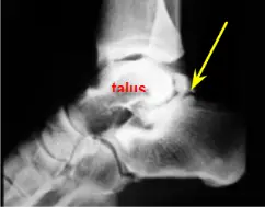
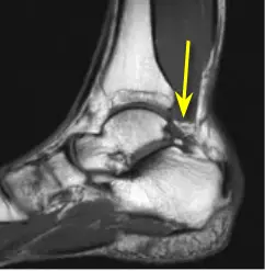
A true os trigonum is a secondary ossification center that has not fully fused with the talus and is connected by fibrous or cartilaginous tissue. (yellow arrow) Frequently it is incorrectly described as a fracture of Stieda’s process which is an enlargement of the posterior process of the talus which is the foot bone that makes up the ankle joint, along with the tibia and fibula bones. This accessory bone lies in the posterior or back part of the ankle.
|
It is estimated that this accessory bone or ossicle occurs in 7-9 percent of the population and by and large it is asymptomatic.
Injury can occur to this bone when the foot is forced into severe plantarflexion (where the foot is bent down and away from the body). This can occur in sports or in dance such as point in ballet. This injury can also occur in repetitive lesser traumatic plantarflexion of the foot. An example would be someone who climbs up and down flights of stairs carrying heavy weights such as furniture movers or delivery men.
mechanism of injury
As the foot is being forced into plantarflexion the posterior process of the talus (Steida's process) as well as the os trigonum (depending on the pathology) is pressed against one of the tendons running in the back of the ankle (flexor hallucis tendon) causing excessive pressure and further exacerbated by the pressure of the posterior part of the ankle, thus resulting in irritation of the Steida's process or the os trigonum.
making the diagnosis of ostrigonum syndrome
When seen in the office the doctor will attempt to reproduce this motion by plantarflexing the foot. If in doing so the patient experiences pain, the doctor should be suspicious of an irritation of the os trigonum or perhaps a fractured posterior talus .
Standard x-rays should differentiate between a fractured talar process or os trigonum syndrome. Typically x-rays are taken of both feet for comparison. When a clear diagnosis is difficult, an MRI may be ordered for better clarity.
|
Symptoms of os trigonum syndrome include a deep pain in the back of the ankle aggravated mostly when the foot is plantarflexed (toes bending downwards). An example would be walking down steep stairs or performing point in ballet. There may also be swelling and tenderness in the back of the ankle. This condition may be mimicked by fracture of Steida's process, as described, as well as achilles tendonitis or ankle sprain.
TREATMENT OF OSTRIGONUM
Conservative care is the treatment of choice in these situations and there is a very high success rate of os trigonum syndrome.
- immobilization with the use of a below knee cast or a CAM walker that does not allow bending, particularly plantarflexion of the ankle. The cast must be worn at all times while the patient is walking. It may be removed for bathing and to ice the area. Crutches may or may not be needed. This regimen is used for 4-6 weeks.
- Along with immobilization the patient is usually put on a regimen of anti-inflammatory medication (if they can tolerate it).
- If after four to six weeks the patient is still having pain then a cortisone injection should be entertained. A second injection may be performed 7-10 days later. Additionally, the patient should continue to wear the cast.
- Once the pain has resolved the patient may need some physical therapy in order to increase range of motion in the ankle, which may have stiffened up due to immobilization. In order to prevent recurrence an ankle brace should be worn to prevent excessive plantarflexion.
Surgical intervention should be considered in those individuals who do not respond to conservative care or those who seem prone to constant recurrence. This extra bone or in the case of Steida's process is not needed for everyday function. The procedure is done as an out patient and requires removal of either the accessory bone or the removal of the broken Steida's process. The patient will have to wear a cast for another few weeks during the healing process. The major complication of this procedure is irritation to the sural nerve which is a sensory nerve that runs down the posterior lateral side of the ankle and down the lateral side of the foot. This may result in sensory loss on the lateral side of the ankle joint and foot which can lead to disability.
REFERENCES
American Academy of Pediatrics
Want more information? CLICK HERE


Recent Articles
-
Vitamin D impact on health
Feb 06, 23 07:17 PM
Researchers are suggesting that the effectiveness of Vitamin D in fighting and preventing disease is predicated on a persons body mass index (BMI). The thinner the person the greater the positive impa… -
Foods to speed up healing
Feb 01, 23 02:41 PM
One of the best ways to help yourself heal faster after surgery is to eat well. Getting the proper nutrition will provide your body with the essentials it needs to promote healing. Here is a suggestio… -
Cancer and Type 2 Diabetes
Jan 25, 23 04:52 PM
An article revealing that older type 2 diabetics have a higher incidence of cancer then non-diabetics. It is suggested that cancer may surpass CVD as the number one cause of death in older diabetics. -
Does glucosamine or MSM reduce arthritis pain?
Jan 22, 23 01:41 PM
A good review of the possible benefits to taking glucosamine, chondroitin or MSM for arthritis. Always beware of the possible side effects of over the counter supplements. -
shin splints
Jan 18, 23 05:12 PM
A great review on the various causes of shin splints, along with treatment options. -
Whats new in skin cancer?
Jan 15, 23 08:32 PM
A presentation of newer skin protection combinations in an effort to better protect the skin from the hazards of sun exposure. -
Causes and risk factors of warts
Jan 14, 23 05:02 PM
A good review of the causes of warts and protective measures you can take to prevent developing them. -
Do chronic wounds need to be dressed daily?
Jan 11, 23 02:18 PM
Because of supply chain shortages as well as staffing shortages particularly during the pandemic, many institutions extended the time between dressing changes for chronic wounds. Is this really the be… -
Food choices that raise your risk of type 2 diabetes
Jan 08, 23 10:07 AM
A good review of how blood sugars can become elevated and the harm that can do. Certain food groups have a tendency to raise your blood sugars and should be avoided. -
Outcome stats from Scarf bunionectomy
Jan 03, 23 03:04 PM
The Journal of Foot and Ankle Surgery recently reported a meta analysis of outcomes in 1583 Scarf bunionectomies that met their inclusion criteria. Adverse events did not seem to be any better or wors…