Heel pain including plantar fasciitis and bone spurs are one of the most common complaints seen in the foot. Plantar fasciitis is an inflammation of the large ligament on the bottom of the foot. Although most cases of plantar fasciitis occur near the heel, often described as sharp heel pain, this condition can be evident anywhere from the heel all the way to the ball of the foot.
In order to feel the plantar fascial ligament, with one hand bend the big toe upwards, run your finger from your other hand along the bottom of the foot, you will feel a large cord like structure that runs from the ball of your foot to your heel. The purpose of this structure is to act like a bowstring in order to support the structure of your arch.
In the picture below, the yellow band represents the plantar fascial ligament. Notices how it attaches from the heel to the ball of the foot.
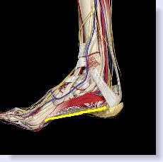
Factors such as foot structure, excessive body weight, occupations that require a lot of standing especially on concrete floors and physical activity such as participating in athletic activities can all lead to excessive stretching and inflammation resulting in plantar fasciitis type of heel pain.
The problem with most foot symptoms unlike a hand injury for example, is that it is very hard to “rest” a foot in order to allow it to get better. So every time you take a step you are reinjuring an injured area and for that reason foot injuries can take a long time to heal, especially heel pain because every time you take a step you are putting pressure on the heel.
Most people relate a similar story in that they find they have the most pain on initial ambulation such as getting out of bed in the morning or getting up from a chair after being seated for a long period of time. As they begin to walk around the pain will usually subside, to varying degrees in different people. However, many people will report that as they begin to do a lot of walking the heel will start to hurt even more.
In my experience, I have found that plantar fasciitis rarely occurs alone. When there is excessive inflammation particularly at the insertion of the ligament into the heel bone, calcification can occur and you end up with what is routinely known as a heel spur which is the classic bone spur in the foot, (although there are other types of bone spurs that occur in other parts of the foot). One could argue that this is simply an exacerbation of the plantar fasciitis.
Below is a picture of a true heel spur; a calcification of the plantar fascial ligament.
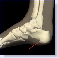
Additionally, when the pain in the heel seems to worsen the more you ambulate other problems may be present. In addition to the plantar fasciitis you may also be suffering from a heel bursitis or even a heel neuroma, which is simply a pinched nerve that gets entrapped in the area of inflammation. Another problem that can add to the heel pain is lack of fat in the heel. The fat is supposed to act as a cushion, but some people either do not have enough fat, or as we age we lose some of the fat, and now the heel bone becomes bruised because there is not enough fat to protect it. It is essential that a well-trained foot specialist be consulted to rule out the cause of the heel pain. (These are the most common but not an all inclusive list of causes of heel pain).
Making the right diagnosis and eliminating the factors aggravating the heel pain are essential in alleviating the problem. If my patient happens to be very much overweight or wears very flimsy shoes, the chances of success in alleviating the problem are greatly diminished unless those issues are addressed.
There are a number of ways to treat plantar fasciitis and heel spurs. Because of this, no one treatment will work for everyone. Treatment options include short term oral anti-inflammatory medication. Cortisone injections can be helpful, usually done in a series of three over a three-week period, especially if you suspect an associated bursitis. Physical therapy can be helpful. If I feel that there is also a neuroma, or pinched nerve involved in the mix, I will usually begin a series of denatured alcohol injections.
Depending on the cause of heel pain, my favorite treatment is the use of orthotics. An orthotic is a device that fits in your shoes and supports the arch and plantar fascial ligament in an effort to keep it from overstretching when you walk. As far as I am concerned, an orthotic has to be the cornerstone of any treatment for heel pain. Unless you can "rest" the plantar fascial ligament, with the use of an orthotic, the ligament will continue to be over stretched and thus continually irritated and will only delay the healing process. I use both custom-made prescription devices and over the counter type orthotics. {see my recommended orthotic} and click here for more information on orthotics for heel pain.
Looking for an effective, safe means to reduce or eliminate your foot pain, consider BLU-MJK. Use it in place of oral anti-inflammatory medication. This is topical medication that you just rub into the painful area. Click the picture below
During the night the plantar fascial ligament will tighten up from lack of use. The first steps that people take in the morning can be very painful as their bodyweight stretches out the ligament. The use of a night splint can be helpful in certain individuals to keep the ligament from tightening up and thus making those initial steps in the morning far less painful. If you are one of those individuals who has excruciating pain getting up in the morning than I would strongly suggest considering a night splint. It should dramatically improve your situation and will also be very helpful in eradicating your heel pain. My recommended night splint is the Ossur/Royce Medical Airform Night Splint..
Another type of heel pain may be caused by nothing more than a bone bruise and actually have nothing to do with the plantar fascial ligament that we have been discussing. The one part of the body where we need to have fat, is the heel. In this case the fat works to cushion the heel bone. As we age, the one part of the body where we actually lose fat is on the bottom of the foot. In these individuals since there is little fat under the heel bone, it becomes bruised from constant walking. It should also be pointed out that some younger individuals do not have much fat under the heel as well.
You can perform a simple test to see if this is the source of your heel pain. Press directly under the heel bone with your finger. If you can actually feel the bone and there is pain upon pressure and this pain is not evident as you move your finger a little bit forward, toward the toes, then you may have a bone bruise. Next, with your other hand, place it around the heel encompassing the outside, back and inside portion of the heel. Squeeze slightly, which will force whatever fat you have back under the heel. Now, with your other hand, press the bottom of the heel bone again. If you notice less pain with the heel cupped and the fat underneath, compared to when the fat was not pushed back under the heel, then there is a very good chance you have a bone bruise.
The best treatment I have found for this condition is through the use of a hard plastic heel cup that sort of looks like an egg shell. Placing this heel cup on your heel either directly under or over your sock or stocking forces whatever fat you have in the heel area back underneath the heel to cushion the heel bone. This will generally work far better than an orthotic or even a gel type heel cup.
When conservative therapies fail to alleviate the problem, more aggressive treatments are called upon. I have found, and the literature will bear me out that 90-95% of heel pain sufferers will respond to the conservative treatments I have discussed.
If you have reached the point where conservative measures have failed and are thinking of the more aggressive therapies I would suggest you have an MRI of the heal prior to consenting to those treatments. Other less common causes of heel pain include stress fractures of the heel bone as well as possible tears in the plantar fascial ligament, along with ruptures of the adjacent muscles attaching into the heel. Sometimes a bone cyst may be present in the heel bone as well. These should be evident in the MRI.
Within the last couple of years many who treat heel pain have started to make a distinction between plantar fasciitis, which we have been discussing and what is known as plantar fasciosis. Plantar fasciitis represents an inflammation on the bottom of the heel for which the various treatments we have already discussed, are indicated, in an effort to quiet down the inflammation and reduce pain.
The latest trend in treating heel pain is making a distinction based on long you have been suffering from your heel pain. If your heel pain has been present for a minimum of two to three months you may actually be experiencing plantar fasciosis. In this condition the pain may actually be due to scarring and fibrosis on the bottom of the heel. Ligaments as well as tendons are poorly vascularized, meaning they tend to get most of their blood supply from surrounding tissue as they do not have enough of their own blood supply. So, what happens in plantar fasciosis is you are left with chronic pain, the body is not attempting to heal itself and all the previous treatments you have received, at best, are giving you temporary relief. Once a diagnosis of plantar fasciosis is made treatment is aimed at improving the blood supply to the heel ligament in an effort to promote healing as it is blood that brings nutrients to an injured area in order to make it well.
Some of the newer treatments being used to increase blood flow to the area are listed below.
A relatively new treatment option is known as extracorporeal shock wave therapy. The science behind this is the same theory used to break up kidney stones. It is high power ultrasound directed at the site of pain. There is strict FDA regulations as to when it is indicated; basically all other options other than surgery have failed. In my experience I have seen some very dramatic results but I also must report that some patients have been no better off after the procedure. It should also be noted that the procedure is covered by some but not all insurance companies and that it can be costly if you have to pay for it out of your own pocket. Click here for more detailed information on shock wave therapy.
Another option is known as low impact shock wave therapy. Whereas the previously stated high impact shock wave is very expensive and does require anesthesia, low impact shock wave therapy is far less expensive (because the machinery is less expensive), does not usually require anesthesia, but may require 3-5 treatment sessions in order to work. It too is a non-invasive therapy like high power shock wave therapy and is usually performed right in the office.
A second option for plantar fasciosis is platelet-rich plasma therapy (PRP). The patient's blood is drawn and spun down creating a small amount of platelet rich material. PRP contains growth factors and bioactive proteins that influence the healing of tendon, ligament, muscle, and bone. This material is then injected into the heel at the site of most pain. Some doctors will inject it under ultrasound guidance. There is a often a short lived pain experience by the patient due to the amount of inflammation that is created. That and the fact that blood has to be drawn and that an injection into the heel has to occur are the only real downsides to the treatment. Since your own blood is being injected, there is no problem with cross contamination. Once again the idea here is to re-create inflammation by the introduction of a high concentration of platelets in order to get the healing process going once again. Because we are looking to produce inflammation, the use of anti-inflammatory medication would be contra-indicated after the procedure.
The third option for plantar fasciosis is what is known as Topaz Coblation Therapy. This is an invasive procedure that is usually carried out in the operating room under sedation. In this procedure a number of small holes are made in the area of the most pain; a needle is inserted into each hole and an electrical charge is given off. The electrical charge results in microscopic cutting of the plantar fascial ligament, an increase in blood flow (inflammation) and break up of the scar tissue that may be present. Even though this is a relatively minor invasive procedure there will be some down time following the procedure as recovery can be slow and somewhat painful.
Another new procedure based on the same principles as the one above is known as Dry Needling whereby anesthesia is first infiltrated into the heel area. Then using ultrasound guidance, an empty hypodermic needle is inserted into the plantar fascial ligament in the area giving the patient the most pain. This is repeated multiple times. The theory here, once again, is to create blood flow into a structure that is generally poorly vascularized. The increased blood flow of course then brings nutrients into the area to heal the plantar fascial ligament, just like blood normally does for other injured parts of the body.
It is also felt that the actual needling of the plantar fascial ligament will also help break up any scar tissue that may have formed. The authors of the original study also recommended directing a cortisone injection into the area once the dry needling was finished to reduce inflammation. In my opinion I do not think the cortisone injection is very helpful for a couple of reasons. One, more than likely patients who are undergoing this procedure have already had one or more cortisone injectons (which did not work) and for anyone who treats plantar fasciitis, it is not very difficult to direct the cortisone into the painful area even without ultrasound guidance. Two, one of the theories regarding recalcitrant heel pain is that the brain is not recognizing the inflamed plantar fascial ligament and thus is not doing what needs to be done to fight the inflammation. This is part of theory already mentioned above regarding the concept of plantar fasciosis.
So, in my mind I would be more inclined to avoid the cortisone injection and allow the inflammation to happen.
After the office procedure it is recommended that the patient wear a walking boot for one week to protect the plantar fascial ligament because since it has been punctured in multiple areas this may actually weaken the ligament and there is a chance of rupture. After the boot is removed I would recommend wearing a good orthotic for a couple more weeks.
The benefits of this procedure is that the authors of this study suggest a 95% success rate, but please keep in mind this was done on a small population of people with heel pain. They also found that relief lasts upwards of ten months and probably longer is the patient takes the necessary steps to avoid re-injury. Relief may not be immediate, but should occur within two to three weeks. This treatment is a one time treatment, performed in the office and since it is not a surgical procedure there is overall less risk and less expense.
Lastly, when all else fails, surgical intervention should be entertained. Today’s foot specialist is trained in doing both conventional large incision procedures and minimal incision procedures. Keep in mind, that surgery for a heel spur or plantar fasciitis can have a very long recovery period. The simple truth of the matter is that after surgery, every time you step on that heel you are basically aggravating the surgical site and for that reason the healing process can take a long time. Thankfully, most cases of heel pain can be resolved without surgical intervention.
Update, November 2010 Like any surgical procedure there are inherent potential complications. One of the problems with any surgery on the plantar fascial ligament is it that will tend to weaken the function of that ligament. At a medical conference I just attended we were shown a before xray prior to a partial plantar fascial release and then an xray taken ten years later revealing a marked drop in the arch of the foot. Now, even though the patient was not having any problems with his foot, the fact that his arch had collapsed so much would lead one to believe that he will eventually suffer from early arthritic changes within the foot as a result of the change.
posterior heel pain
The second area where people experience heel pain is on the back of the heel. Although there can be a connection between pain on the bottom and back of the heel, the causes of posterior heel pain generally differ from that of plantar heel pain.
The most common cause of posterior heel pain that I see is generally referred to as pump bump. Typically a woman will present to the office complaining of a sharp pain on the back of the heel just slightly off the center. There will be no history of trauma, although the patient may be required to do a lot of walking in a dress shoe. The pain will be at its worst when in dress shoes, may be less painful in sneakers and may be hardly noticeable when barefoot.
Examination of the back of the heel will reveal a slightly enlarged area that may be slightly red in color and usually very painful to touch. Not coincidentally, you will notice that the top of the patient’s dress shoe just happens to cut across the heel at that level. What has happened is that through constant wearing of dress shoes, the shoe itself has irritated the underlying bone, the bone has slightly enlarged as a result of that irritation and in an effort to protect the area of irritation, the body has formed a bursal sac which itself has become inflamed and is now a bursitis.
Below is the usual site for a ‘pump bump’ or bone spur with bursitis.
The obvious treatment would be to avoid wearing the shoes that aggravate the problem; that is easier said than done. I have been in this business long enough to know that a fashionable woman is going to continue to wear the shoes that look the best even though they might not feel the best.
That said, sometimes adding a small heel lift to the shoe will help as it may raise the heel just enough so that the top back portion of the shoe does irritate the enlarged bone and bursal sac. If the area is not too inflamed and painful, one may try some of the new gel cushions that are available in an effort to reduce friction.
From a medical stand point, short term anti-inflammatory medication may help but of course will not cure if the shoe is still irritating the area. Cortisone injections as well as physical therapy may also be effective.
In instances where conservative measures fail, the patient should entertain surgical excision whereby the overgrowth of bone is removed as well as the overlying bursitis. This is usually reserved for people who have stopped wearing the shoes that originally caused the problem, but are still having pain nonetheless. Unfortunately, many foot problems can continue to be bothersome even when the causative agent is identified and removed. (Once you have it; you have it!)
The other type of posterior heel pain that we see involves irritation of the Achilles tendon. The cause of this irritation may be due to trauma directly to the tendon, overuse of the tendon which is particularly a problem in men over the age of 40 who continue to play a lot of sports. People who have a tight heel cord, whereby it is very difficult to raise the foot at the level of the ankle, are more prone to Achilles tendon problems.
Below is a picture of the Achilles tendon.
The patient will present with tenderness and possibly swelling in the area of Achilles tendon anywhere from where it attaches into the heel bone to just above the ankle. An xray may also reveal the formation of bone spurs at the level where the tendon attaches into the heel. The spurs can become painful if they are irritated enough. Further complicating the situation is the fact that there is a natural occurring bursal sac that sits between the heel bone and Achilles tendon which may also become inflamed and painful in the process.
If there has been trauma to the area it is important make sure all or part of the tendon has not torn. You should see a doctor right away, even though you may be able to walk, you may still have a partial tear. A torn Achilles tendon is a very debilitating condition.
Icing the area for 24 hours can be very helpful as well as anti-inflammatory medication. Rest would also be advised. I am not big on ice after 24 hours; I generally switch my patients to heat in an effort to increase blood flow to the area to enhance the healing.
If the Achilles tendon has been strained rather than traumatized, the above applications apply. Adding a one quarter inch heel lift can be of benefit because it will shorten the tension on the heel cord especially in those who exhibit a tight Achilles tendon. If the stain is severe enough immobilization of the ankle may be necessary.
FIND A PODIATRIST
Are you in need of a podiatrist in order to solve your foot or ankle problem. Click Here to locate a podiatrist in your area.