scars
AUTHOR: Marc Mitnick DPMREVIEWED BY: Podiatric Medical Review Board
home --> scars
WHAT IS A SCAR
Also known as cicatrix is a natural part of healing. Scarring will occur as a result of trauma or surgical incision. Most organs of the body will exhibit a degree of scarring, but the focus of this discussion is how the skin is affected.
During the phases of wound repair of which there are three, the final phase, or remodeling phase, healthy skin is replaced with a more fibrous type skin in an effort to bridge any defects seen in the skin. In the majority of instances the healing tissue is probably not even visible nor does it have any impact on the affected area. The resulting remodeling is usually flat and over time becomes less noticeable.
factors that affect scarring
The degree of scarring that may occur depends on:
- a person's age. This problem tends to form more predominantly in younger individuals.
- sex
- heredity
- nutritional status of the person
- the severity of the original wound
types of abnormal scars
|
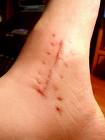
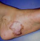
Most troublesome cicatrix are classified into two groups. One is a hypertrophic scar which is usually a large red scar that forms within the confines of the original wound. They are very vascular which gives rise to their red appearance. They tend to be inelastic in nature which can cause pain. Sometimes in spite of their original size the can fade over time in many cases.
The other type is a keloid which are also raised, red, but tend to cover skin beyond the boundaries of the original wound. More importantly these do not tend to fade over time. Even when surgically excised, they tend to recur. It is thought that keloids have a genetic disposition.
|
We should also mention that there is another type of scarring that is seen typically as a result of burns. Burns cause a contracture type of scarring where the affected area becomes extremely inelastic and can be quite uncomfortable, depending on the size of the lesion.
Of course cicatrix can appear any where on the body, but this discussion will be limited to the foot and ankle. Excessive scarring particularly in an area over the front of the ankle or perhaps the big toe joint can result in a limitation of motion due to the inability of the lesion to "stretch" like healthy skin. Many times this can be due to poor skin closure of the skin by a surgeon, sometimes trauma to that area will have a similar result.
foot surgery and the risk of scars
One of the biggest problems we as foot surgeons face is surgical intervention on the bottom of the foot. The skin of the bottom of the foot is generally thicker than skin elsewhere plus it is prone to more tension as the result of bearing weight on the foot. Typically when planning a surgical procedure, great care is taken as to where the incisions are going to be placed in an effort to reduce the stress on the incision. It is not uncommon to keep our patients non-weight bearing for a few weeks after the procedure just to keep tension off the wound and reduce the chance of scarring. Many times additional sutures will be placed in the wound in an effort to reduce tension, however, the down side to that is if the patient has a reaction to the suture material itself, having excess sutures in the wound may actually exacerbate the scar problem.
A perfect example of avoiding plantar incisions is in the surgical excision of Morton's neuroma. The involved nerve actually runs on the bottom of the foot but in almost all instances incisions are made through the top of the foot, to get to the nerve, and avoid scarring on the bottom of the foot.
Weight bearing sections of the foot, particularly the ball of the foot are very prone to scarring as a result of trauma or incision. When patient's have a benign growth on the ball of the foot, such as a warts or porokeratosis. they should not be too quick to allow their doctor to surgically excise or hyfrecate (burn them out) these lesions as an end result of scarring may ultimately be more uncomfortable than the actual growth itself. More conservative means should be used to eradicate the problem. If a suspected malignancy is present then there is really no choice, however, I would suggest something less traumatic like a needle biopsy to first assess malignancy before actually excising the whole lesion.
TREATMENT OF SCARS
As time passes more and more treatments for this problem have come to marketplace. Not all the products available today have been adequately tested to really know their true effectiveness.
Surgical revision of the skin overgrowth has been around for ages. Depending on the size and type of cicatrix there are two different approaches. One is to excise the lesion and re-suture the area. The surgical approach is to perform plastic surgery procedures on the area such as what is known as Z-plasty or W-plasty which are procedures that actually redirect the scar. This is usually done on lesions that are limiting motion. There is however, a reasonably high incidence of recurrence somewhere near the fifty percent range.
Cortisone injections for the reduction of skin overgrowth has been around for years. Used with other treatments such as surgical excision, the injections may be given every two weeks in an effort to reduce the inflammatory response of the wound healing phase.
Radiotherapy has been used for larger keloids but it is important that the patient knows there is some evidence that it may increase carcinogenic activity in the surrounding area.
Laser therapy and dermabrasion has been used to "smooth" down scars but in recent years seems to becoming a less attractive option due to the high recurrence rate and the fact that it can take months to finally heal and during this time period the scars can be painful, swollen and red.
Cryotherapy or the use of liquid nitrogen works by freezing the scar and thus causes cell damage which prevents an over production of fibrous cells. Treatment can be performed every three to four weeks until your doctor is satisfied with your progress. This procedure may also tend to be painful and in some cases cause a depigmentation (loss of color) in the skin.
There are a few skin creams on the market which claim to reduce scarring. They can make a difference in the appearance of the scar but it is a cosmetic improvement, not an actual thinning of the skin overgrowth. One such product is Mederma.
Compression therapy, whereby continuous pressure on a scar may actually thin it out over time. It does so by suppressing the inflammatory phase. Treatment may take upwards of a year depending on the size and location of the scar.
Silicone gels have become popular in recent years. Their method of action is by being placed directly over the scar they create a moist, occluded environment which prohibits increased blood vessel growth which then reduces the inflammatory over response seen in scars. These gel pads are simply taped on the offending skin. The gel sheets may be washed, they happen to be impermeable to bacteria, but over time they will lose their efficacy and will have to be replaced.
REFERENCES
American Society for Dermatological Surgery
American Academy of Dermatology
Want more information? CLICK HERE


Recent Articles
-
Vitamin D impact on health
Feb 06, 23 07:17 PM
Researchers are suggesting that the effectiveness of Vitamin D in fighting and preventing disease is predicated on a persons body mass index (BMI). The thinner the person the greater the positive impa… -
Foods to speed up healing
Feb 01, 23 02:41 PM
One of the best ways to help yourself heal faster after surgery is to eat well. Getting the proper nutrition will provide your body with the essentials it needs to promote healing. Here is a suggestio… -
Cancer and Type 2 Diabetes
Jan 25, 23 04:52 PM
An article revealing that older type 2 diabetics have a higher incidence of cancer then non-diabetics. It is suggested that cancer may surpass CVD as the number one cause of death in older diabetics. -
Does glucosamine or MSM reduce arthritis pain?
Jan 22, 23 01:41 PM
A good review of the possible benefits to taking glucosamine, chondroitin or MSM for arthritis. Always beware of the possible side effects of over the counter supplements. -
shin splints
Jan 18, 23 05:12 PM
A great review on the various causes of shin splints, along with treatment options. -
Whats new in skin cancer?
Jan 15, 23 08:32 PM
A presentation of newer skin protection combinations in an effort to better protect the skin from the hazards of sun exposure. -
Causes and risk factors of warts
Jan 14, 23 05:02 PM
A good review of the causes of warts and protective measures you can take to prevent developing them. -
Do chronic wounds need to be dressed daily?
Jan 11, 23 02:18 PM
Because of supply chain shortages as well as staffing shortages particularly during the pandemic, many institutions extended the time between dressing changes for chronic wounds. Is this really the be… -
Food choices that raise your risk of type 2 diabetes
Jan 08, 23 10:07 AM
A good review of how blood sugars can become elevated and the harm that can do. Certain food groups have a tendency to raise your blood sugars and should be avoided. -
Outcome stats from Scarf bunionectomy
Jan 03, 23 03:04 PM
The Journal of Foot and Ankle Surgery recently reported a meta analysis of outcomes in 1583 Scarf bunionectomies that met their inclusion criteria. Adverse events did not seem to be any better or wors…