melanoma
AUTHOR: Marc Mitnick DPMREVIEWED BY: Podiatric Medical Review Board
home --> melanoma
WHAT IS A MELANOMA
This is a mole that has undergone malignant changes. A malignant melanoma is the most deadly of all skin cancers. A mole is medically known as nevi. Normal moles are usually uniform in color being brown, black or tan. They will have even borders and usually no larger than the diameter of a pencil’s eraser. Moles may change in appearance over time, some are known to even disappear.
Some moles may become larger over time, may develop a mixture of color or may develop uneven borders. These moles are known as dysplastic nevi and are more prone to becoming malignant.
People at high risk for developing melanoma are those who have:
- A family history of or have had a melanoma in the past.
- Unusual moles on the skin, or changing moles.
- Fair skin, light hair and eye color, who sunburn easily or tan with difficulty.
- A history of painful or blistering sunburns as children or teenagers.
Since skin cancers are known to develop more commonly in the area of “BANS”, back, arms, neck and scalp which are the areas most exposed to the sun you might be wondering why I am discussing this condition since this web site is devoted to foot problems and the feet are generally not exposed as much to the sun as other parts of the body.
It is precisely for that reason that I am discussing this condition; to make my readers aware that they can occur on the feet and the feet themselves have certain characteristics that may promote the development of this malignancy.
The incidence of melanoma is equal between men and women, but in the lower extremity it is more common in females than males. The majority of cases present between 30-60 years of age.
melanoma facts
There are four different categories of this malignant mole, some of which are more virulent than others. It is not the purpose of this discussion to make you an expert on this condition, but rather make you aware of changes in moles that need to be examined by experts.
TWO UNIQUE LOCATIONS OF MELANOMA IN THE FOOT
Bottom of the foot or in between the toes
The reason we worry about moles in these locations is because of the friction that occurs on the skin in these areas, any moles that develop are more prone to undergoing malignant changes due to that excessive friction. So, just because a mole grows in between your toes and you figure it could not have possibly been affected by sun light is no reason not to keep an eye on it or have it inspected by a doctor.

|
Melanoma under nail
Any dark discoloration underneath the nail whose origin cannot be explained needs to be examined. In the vast majority of the cases the discoloration that may occur under the nail is either from trauma to the nail and the subsequent bleeding that occurs or from a fungus growth that may develop. See my discussion on nail fungus.In both those cases, the discoloration should grow forward as the nail grows and eventually will be cut away at the end of the nail. If there is a dark discoloration under the nail that does not seem to be growing forward, (it just stays in the same place) than that discoloration should be considered suspicious. In my office in most cases I can cut away enough nail to visually examine the nail bed and in most cases the discoloration can be removed leaving a healthy nail bed. If I cannot get to the discoloration that way, then I will anesthetize the toe and remove the whole nail, examine the discoloration and if necessary will biopsy the lesion. Again, in the vast majority of cases the discoloration is either from trauma (dried blood) or from fungus.
Regular self exams, all over your body is the best way to become familiar with the many moles and spots you may have on your skin.
WHAT DOES MELANOMA LOOK LIKE
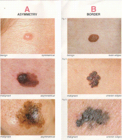
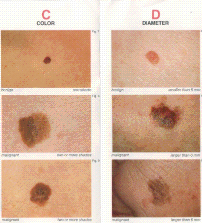
In self examination of your moles you need to inspect them for certain characteristics according to their size, color, shape and edges. These are known as the A-B-C-D’s of self examination. They stand for Asymmetry, Border, Color and Diameter.
melanoma pictures
Below are pictures of various types of moles characterized by the A-B-C-D descriptions. The top mole in each description is normal and the bottom two are malignant.
|
|
ASSYMMETRICAL meaning a line drawn thru them will not create equal matching halves. It is easy to see what this means by the examples above.
BORDER meaning that normal moles have even borders, but malignant moles frequently have uneven borders.
COLOR of brown, black or tan is normal but once a mole starts having different shades of color it becomes suspicious.
DIAMETER of common moles are usually the size of a pencil eraser head in diameter. Early malignant moles tend to be larger than that.
To further complicate the diagnosis of this malignant mole, there is a variant known as amelanotic melanoma. This is a melanoma with no pigment meaning it does not have a dark discoloration to it. Because of their lack of color these lesions are frequently diagnosed later in their growth meaning they tend to be deeper and more invasive. Fortunately, they do represent less than 5% of all melanomas.
They may be misdiagnosed as eczema, granulation tissue and paronychia (infected ingrown nail).
Typically, on the foot, amelanotic melanoma appears as a non-healing ingrown nail. Many paronychias are associated with a pyogenic granuloma which is a section of inflamed skin adjacent to the ingrown nail. These benign lesions are of a vascular makeup and tend to bleed profusely if cut. In most instances your doctor will cauterize the growth after he removes the offending ingrown nail. If the ingrown nail clears up, but the granuloma persists it should be brought to the attention of your doctor for possible biopsy.
When appearing on the skin, amelanotic malignant mole will present as a pink, reddish or even flesh colored. The diagnosis is generally not made until the lesion begins to grow or begins to itch or becomes painful or even begins to bleed. In many instances these lesions will be irritated from chronic shoe or clothing irritation.
As stated before it is important to examine all your moles on your body. Dermatologists recommend yearly examinations.
Interestingly, this year a study out of Rome, Italy found that people with higher serum levels of coenzyme Q (coq 10) has less incidence of melanoma and less metatastasis. (Journal American Academy of Dermatology, 2006 (ISSN:1097-6787)
As a last comment just because a mole somewhere on your body appears uncharacteristic it does not mean it is malignant but what it does mean is that you should have your doctor look at it immediately. A biopsy is the only way to determine the identity of the mole.
REFERENCES
American American Academy of Dermatology
Want more information? CLICK HERE


Recent Articles
-
Vitamin D impact on health
Feb 06, 23 07:17 PM
Researchers are suggesting that the effectiveness of Vitamin D in fighting and preventing disease is predicated on a persons body mass index (BMI). The thinner the person the greater the positive impa… -
Foods to speed up healing
Feb 01, 23 02:41 PM
One of the best ways to help yourself heal faster after surgery is to eat well. Getting the proper nutrition will provide your body with the essentials it needs to promote healing. Here is a suggestio… -
Cancer and Type 2 Diabetes
Jan 25, 23 04:52 PM
An article revealing that older type 2 diabetics have a higher incidence of cancer then non-diabetics. It is suggested that cancer may surpass CVD as the number one cause of death in older diabetics. -
Does glucosamine or MSM reduce arthritis pain?
Jan 22, 23 01:41 PM
A good review of the possible benefits to taking glucosamine, chondroitin or MSM for arthritis. Always beware of the possible side effects of over the counter supplements. -
shin splints
Jan 18, 23 05:12 PM
A great review on the various causes of shin splints, along with treatment options. -
Whats new in skin cancer?
Jan 15, 23 08:32 PM
A presentation of newer skin protection combinations in an effort to better protect the skin from the hazards of sun exposure. -
Causes and risk factors of warts
Jan 14, 23 05:02 PM
A good review of the causes of warts and protective measures you can take to prevent developing them. -
Do chronic wounds need to be dressed daily?
Jan 11, 23 02:18 PM
Because of supply chain shortages as well as staffing shortages particularly during the pandemic, many institutions extended the time between dressing changes for chronic wounds. Is this really the be… -
Food choices that raise your risk of type 2 diabetes
Jan 08, 23 10:07 AM
A good review of how blood sugars can become elevated and the harm that can do. Certain food groups have a tendency to raise your blood sugars and should be avoided. -
Outcome stats from Scarf bunionectomy
Jan 03, 23 03:04 PM
The Journal of Foot and Ankle Surgery recently reported a meta analysis of outcomes in 1583 Scarf bunionectomies that met their inclusion criteria. Adverse events did not seem to be any better or wors…