osteomyelitis
AUTHOR: Marc Mitnick DPM home --> osteomyelitisbone infection
WHAT IS OSTEOMYELITIS
Osteomyelitis is a bone infection which will cause bone destruction. It can occur anywhere in the body but it is especially prevalent in the foot for reasons you will soon come to understand. The condition is caused when bacteria, in most cases, staphylococcus aureus (staph aureus) invades a bone.
types of osteomyelitis
This can occur in three different ways and historically this condition has been classified based on how it occurs.
- Hematogenous osteomyelitis This is a disease of the very young and very old. It occurs from a soft tissue infection elsewhere in the body, that then gets into the bloodstream and travels to a bone somewhere else in the body and invades that bone. In the young it has a greater tendency to invade “long bones” and lodge itself in the most vascular part of the bone known as the metatphyseal region.
In the elderly the most common site is the vertebrae. This type of bone infection is generally not caused by staph aureus but rather by other organisms.
For the sake of completeness this type of bone infection is also caused by IV drug users who basically end up injecting the bacteria into their circulation.
- Contiguous osteomyelitis In this instance the bone becomes infected from an external contaminated source such as penetrating trauma, open fracture, bone surgery or joint replacement. This type can occur at any age and in any bone. This type is very common in the foot.
- Vascular insufficiency osteomyelitis, or poor circulation. This is quite frequently seen in diabetics with diabetic neuropathy. (See my discussion on neuropathy and diabetic foot). Foot ulcers serve as an entrance for infection and bacteria to gain access to the bone by contiguous spread. The general rule of thumb is that if an ulcer fails to heal in six weeks, and assuming has been treated with meticulous wound care, then one should suspect bone infection of the underlying bone.
Note the x-ray below of a right foot. The blue arrow points to the area of bone destruction on the great toe. Notice the mottled appearance of the bone and the irregularities at the edges of the bone. Compare this to the normal bone of the first metatarsal which is noted by the red arrow. For those of you perceptive enough, you will notice the second toe has been amputated.

|
SYMPTOMS OF OSTEOMYELITIS
|
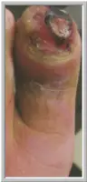
Symptoms associated with bone infection include local signs of pain, swelling and redness in the area of the infection. Note the picture to the left. Systemic signs will include chills, fever and malaise.
As previously stated staph aureus is the most common bacteria associated with this type of infection, but is not the only one. Pseudomonas aeruginosa is also a very common bacteria and this is typically seen as a result of puncture wounds. Diabetics and others with compromised circulation and immune systems may present with this bone infection from multiple strains of bacteria.
Early detection is very important as to limit the destruction of bone and to stand a better chance of resolving the infection with antibiotics. That is where the problem arises for the doctor. This is a disease that can be difficult to detect and there are other conditions which may give a false positive to the tests for osteomyelitis.
DIAGNOSING OSTEOMYELITIS
Since this is a bone infection one would think that an x-ray would show a bone infection. The problem is that many times they are initially inconclusive. Bone has to lose upwards of fifty percent of its density before changes will be seen on x-ray. By the time it takes the bone to lose this much density (2 to 6 weeks) and show up on x-ray the bacteria has fairly well infiltrated the bone.
Many doctors will order a bone scan to make the diagnosis. There are mixed results with this method. Bone scans may be “hot” or positive for conditions other than osteomyelitis such as trauma, (surgical trauma included), fractures, as well as diabetic osteolysis which is bone destruction due to the ravages of diabetes.
An MRI is the most sensitive and specific imaging study for defining bone infection but they are not universally available and they are expensive.
Absolute definitive diagnosis is made based on bone culture and biopsy. Using various techniques depending on location, a piece of the diseased bone is actually removed and cultured for bacteria. It is important that the surgeon probe down to bone to get the culture as opposed to taking soft tissue samples as there can be a difference in organisms found at each site. A patient should also be off all antibiotics for at least 48 hours to allow for the most accurate results.
TREATMENT OF OSTEOMYELITIS
In most cases of bone infection, surgery is the treatment of choice with the exception of the hematogenous variety which many times can be treated successfully with antibiotics for upwards of six weeks. This is usually a combination of parenteral antibiotics (intravenous or injectable) followed by oral antibiotics. In some cases antibiotic beads have been implanted in the infected area is an effort to disperse a continual saturation of antibiotic to the infected bone. However, this modality is still considered controversial. A full course of antibiotics must be taken in order to destroy the bacteria otherwise you run the risk of a chronic infection.
Most other cases will require surgical debridement (removal) of all the diseased bone particularly because some organisms form a gel layer over and in the bone protecting them from the effects of antibiotics. The chief consideration is what will be the resulting function at that level of surgery. Having the end of a toe amputated generally will not hinder a patient in their ability to walk, but more extensive surgery in other parts of the foot may make walking afterward very difficult, so all this has to be planned for prior to surgery. It should also be noted that in cases of surgical debridement, antibiotic therapy will also be necessary.
Acute osteomyelitis that is either not treated or improperly treated will go on to becoming a chronic bone infection. Another scenario is one where the circulation to the extremity is so bad to begin with that surgical intervention would almost certainly lead to a non-healing surgical wound with the potential for loss of limb or even life. In fact may infectious disease experts never consider osteomyelitis as cured but rather as “arrested”. A person can live with a chronic bone infection assuming it does not adversely affect the body part in question. However, chronic disease does have its pitfalls. The infected bone as well as sinus tracts may undergo malignant changes, there may be constant pain in the site and the everyday care of the infected site may cause more problems than it is worth.
REFERENCES
Want more information? CLICK HERE


Recent Articles
-
Vitamin D impact on health
Feb 06, 23 07:17 PM
Researchers are suggesting that the effectiveness of Vitamin D in fighting and preventing disease is predicated on a persons body mass index (BMI). The thinner the person the greater the positive impa… -
Foods to speed up healing
Feb 01, 23 02:41 PM
One of the best ways to help yourself heal faster after surgery is to eat well. Getting the proper nutrition will provide your body with the essentials it needs to promote healing. Here is a suggestio… -
Cancer and Type 2 Diabetes
Jan 25, 23 04:52 PM
An article revealing that older type 2 diabetics have a higher incidence of cancer then non-diabetics. It is suggested that cancer may surpass CVD as the number one cause of death in older diabetics. -
Does glucosamine or MSM reduce arthritis pain?
Jan 22, 23 01:41 PM
A good review of the possible benefits to taking glucosamine, chondroitin or MSM for arthritis. Always beware of the possible side effects of over the counter supplements. -
shin splints
Jan 18, 23 05:12 PM
A great review on the various causes of shin splints, along with treatment options. -
Whats new in skin cancer?
Jan 15, 23 08:32 PM
A presentation of newer skin protection combinations in an effort to better protect the skin from the hazards of sun exposure. -
Causes and risk factors of warts
Jan 14, 23 05:02 PM
A good review of the causes of warts and protective measures you can take to prevent developing them. -
Do chronic wounds need to be dressed daily?
Jan 11, 23 02:18 PM
Because of supply chain shortages as well as staffing shortages particularly during the pandemic, many institutions extended the time between dressing changes for chronic wounds. Is this really the be… -
Food choices that raise your risk of type 2 diabetes
Jan 08, 23 10:07 AM
A good review of how blood sugars can become elevated and the harm that can do. Certain food groups have a tendency to raise your blood sugars and should be avoided. -
Outcome stats from Scarf bunionectomy
Jan 03, 23 03:04 PM
The Journal of Foot and Ankle Surgery recently reported a meta analysis of outcomes in 1583 Scarf bunionectomies that met their inclusion criteria. Adverse events did not seem to be any better or wors…